Thoracolumbar Fracture
What is a thoracolumbar fracture?
A thoracolumbar (thor-ah-ko-LUM-bar) fracture is a break in one or more of your thoracic and lumbar vertebrae. The vertebrae are the bones that make up your spine. The thoracic vertebrae are the 12 bones between your neck and lower back. They are connected to your ribs and help the ribs move when you breathe. The lumbar vertebrae are the five bones between your chest and hips. Lumbar bones are the largest and strongest of all vertebrae.
What causes a thoracolumbar fracture?
Thoracolumbar fractures are usually caused by severe injury to the spine. When the spine is damaged, there may also be damage to the spinal cord. The following are some causes of severe spinal injury:
Car accidents: Children may wear a lap belt, but not wear the shoulder belt when passengers in a vehicle. If a car stops suddenly, too much force may be put on the spine. Twisting or the shear force from the accident may push the thoracic or lumbar bones out of place.
Falling from a great height.
Jumping from a high position and landing on your feet.
Osteoporosis: This is a condition where bones become weaker than normal. It may be seen in older adults and other people.
What are the signs and symptoms of a thoracolumbar fracture?
You may have one or more of the following:
Abnormal curve of the spine.
Bruising and swelling on your back.
Decreased feeling, weakness, or paralysis (loss of movement) of the legs.
Limited movement of your back.
Pain when moving. The pain may not be felt right away and may begin hours after the injury.
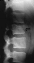
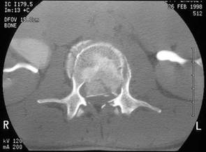
How is a thoracolumbar fracture diagnosed?
You may have one or more of the following:
X-rays: You may need x-rays of the thoracolumbar area to check for broken bones or other problems.
Computerized tomography scan: This is also called a CT or CAT scan. A CT scan is a type of x-ray that uses computers to take pictures of the thoracic and lumbar spine.
Magnetic resonance imaging scan: This is also called an MRI. An MRI uses magnetic waves to take pictures of the thoracic and lumbar spine.
Bone scan: This is a test to look at your bones. You are given a small, safe amount of radioactive dye in an IV. Pictures are then taken of your bones. Caregivers can look at the pictures for broken bones, infections, or cancer in the bones.
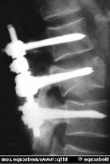
How is a thoracolumbar fracture treated?
Treatment will depend upon which bones were damaged and the kind of fracture you have. Thoracolumbar fractures that are mild may be treated with bed rest. For support, you may need to wear a back brace or have a cast made for your back. Sometimes a corset (binder) may be used to support a weak spine. To decrease the load on a broken spine, you may need to use a walker. A severe thoracolumbar fracture may require surgery to return the bones to their normal position.
Courtesy of: http://www.drugs.com/cg/thoracolumbar-fracture.html
Spinal Cord Injury
Injury to the spinal cord in the neck causes a condition known as tetraplegia or quadriplegia. These terms mean exactly the same thing, one is a Greek term and the other is Latin.
There is injury to the spinal cord between the spinal cord segments C1 and T1
This causes paralysis and loss of feeling involving all 4 limbs as well as the bladder, bowel and sexual organs.

Paraplegia
Injury to the spinal cord below the neck causes paraplegia.
There is injury to the spinal cord below the T1 cord segment.
This causes weakness and loss of feeling in the trunk, legs and bladder, bowel and sex organs. But the arms and hands are normal.
Level of injury
We describe the level of the injury by referring to the last spinal cord segment where the movement and feeling are normal.
For example, if your injury is called “C5” this means that you would be able to bend your elbow normally and would have normal feeling to around the level of your elbow but below that the movement and feeling would be reduced.
Incomplete
An incomplete injury is one in which there is some movement or feeling below the level of the injury or in the genital region.This implies that the damage in the spinal cord does not involve the whole spinal cord and that some messages are getting past the area of damage.
The chances of improvement and recovery are better if the injury is incomplete.

Complete
A complete injury is one in which there is no movement or
feeling in the genital region.
This means that the damage in the spinal cord involves the
whole spinal cord and no messages are getting past the
area of damage.
The chance of improvement and recovery if the injury is
complete is much lower.
if pressure is applied to the spinal cord or
if the blood supply (which brings the oxygen to the cord) is cut off for more than about 15 minutes.
spinal cord damage is probably due to a combination of both factors.

Please refer to the following website: http://www.health.qld.gov.au/qscis/info_handbook.asp