
Radiation Therapy: Brain Tumours (Stereotactic Radiosurgery)
Radiation Therapy of Brain Tumours and Lesions : Stereotactic Radiosurgery

What is stereotactic radiosurgery and how is it used?
Stereotactic radiosurgery is a highly precise form of radiation therapy used primarily to treat tumors and other abnormalities of the brain. Despite its name, stereotactic radiosurgery is a non-surgical procedure that uses highly focused x-rays to treat certain types of tumors, inoperable lesions and as a post-operative treatment to eliminate any leftover tumor tissue.
The treatment involves the delivery of a single high-dose—or sometimes smaller, multiple doses—of radiation beams that converge on the specific area of the brain where the tumor or other abnormality resides. Using a helmet-like device that keeps the head completely still and three-dimensional computer-aided planning software, stereotactic radiosurgery minimizes the amount of radiation to healthy brain tissue.
Stereotactic radiosurgery is an important alternative to invasive surgery, especially for tumors and blood vessel abnormalities located deep within or close to vital areas of the brain. Radiosurgery is used to treat many types of brain tumors, both benign or malignant and primary or metastatic. Additionally, radiosurgery is used to treat arteriovenous malformations (AVMs), a tangle of expanded blood vessels that disrupts normal blood flow in the brain and is the leading cause of stroke in young people.
Although stereotactic radiosurgery is often completed in a one-day session, physicians sometimes recommend a fractionated treatment, in which treatments are given over a period of days or weeks. This is referred to as stereotactic radiotherapy.
Stereotactic radiosurgery works in the same way as other forms of radiation treatment. It does not actually remove the tumor; rather, it distorts the DNA of tumor cells. As a result, these cells lose their ability to reproduce. Following the treatment, benign tumors usually shrink over a period of 18 months to two years. Malignant and metastatic tumors may shrink more rapidly, even within a couple of months. When treated with radiosurgery, arteriovenous malformations (AVMs) begin to thicken and close off.
Who will be involved in this procedure?
The treatment team is comprised of a number of specialized medical professionals, typically including a radiation oncologist, neurosurgeon, medical radiation physicist, dosimetrist, radiation therapist, radiation therapy nurse, and neurologist or neuro-oncologist. The radiation oncologist and neurosurgeon oversee treatment and interpret the results of radiosurgical procedures.
The radiation oncologist, a specially trained physician who heads the treatment team, sets an individualized course of treatment with the help of the medical radiation physicist, who ensures the delivery of the precise radiation dose. A dosimetrist, under the supervision of the physicist, calculates the exposures and beam configurations necessary to deliver the dose prescribed by the radiation oncologist. A highly trained radiation therapist positions the patient on the treatment table and operates the machine. The radiation therapy nurse provides the patient with information about the treatment and possible adverse reactions.
What equipment is used?
There are three basic forms of Stereotactic radiosurgery, each of which uses different instruments and sources of radiation:
Gamma Knife, which uses 201 beams of highly focused gamma rays. Because of its incredible accuracy, the Gamma Knife is ideal for treating small to medium size lesions. See the Gamma Knife page for more information.
Linear accelerator (LINAC) machines, prevalent throughout the world, deliver high-energy x-ray photons or electrons in curving paths around the patient's head. The linear accelerator can perform radiosurgery on larger tumors in a single session or during multiple sessions, which is called fractionated stereotactic radiotherapy. Multiple manufacturers make this type of machine, which have brand names such as Peacock®, X-Knife®, CyberKnife®, Clinac®. See the Linear Accelerator page for more information.
Particle beam (proton) or cyclotron is in limited use in North America. However, several new facilities are being built.
Who operates the equipment?
The multidisciplinary team, including the radiation oncologist, medical physicist and dosimetrist, plan and prescribe the appropriate treatment dose and delivery. The radiation therapist is responsible for operating the radio surgical equipment from a protected area nearby. The radiation therapist can observe the patient through a window or on a closed-circuit television and is able to communicate with the patient throughout the procedure.
Is there any special preparation needed for the procedure?
Prior to the procedure, you may be given a special shampoo with which to wash your hair. You will be asked not to eat or drink anything after midnight on the night before your treatment. You should ask your physician what to do about taking any normal medications on the day of your treatment and bring those medications with you to the procedure. You should also tell your physician if any of the following apply to you:
You are taking medications by mouth or insulin to control diabetes.
You are allergic to intravenous contrast material, shellfish, or iodine.
You have a pacemaker, artificial heart valve, defibrillator, brain aneurysm clips, implanted pumps or chemotherapy
ports, neurostimulators, eye or ear implants, stents, coils or filters.You suffer from claustrophobia.
Stereotactic radiosurgery is usually performed on an outpatient basis. However, be prepared to spend up to 16 hours in the hospital. You will need to have a family member or other support person accompany you, remain with you at the treatment facility, and drive you home afterward.
On treatment day, you will be asked to remove all jewelry, makeup (including nail polish) hairpieces, contact lenses, eyeglasses and dentures. You will be asked to change into a gown for your procedure. An intravenous (IV) line may be inserted into your arm for any necessary medications. You may receive medications to help you relax and to prevent dehydration.

How is the procedure performed?
Stereotactic Radiosurgery Using the Gamma Knife
NB: VERY LIMITED AVAILABILITY IN AUSTRALASIA
Gamma Knife radiosurgery involves four phases: placement of the head frame, imaging of tumor location, computerized dose planning, and radiation delivery.
In the first phase, a box-shaped head frame is attached to your skull using specially designed pins to keep your head from moving until the treatment session is finished. This lightweight aluminum head frame is a guiding device that makes sure the Gamma Knife beams are focused exactly where the treatment is needed.

Next, you will be taken to an imaging area where a computed tomography (CT) scan and/or magnetic resonance imaging (MRI) will be performed to show the exact location of the tumor in relation to the head frame.
During the next phase, you will be able to relax for an hour or two while your treatment team performs a computer-aided treatment plan that will optimally radiate the tumor.

Next, you will lie down on the Gamma Knife bed where your physician will describe the number and length of treatments to expect. Your head frame will then be attached to a helmet that has several hundred holes in it to allow individual rays of radiation to target specific areas of the brain.
The treatment team will then go to another room so that your treatment can begin. You will be able to talk to your physician through a microphone in the helmet and a camera will allow the team to see you at all times. The bed you are lying on will move backward into the treatment area. You may hear a chime at this point and a click as the helmet locks into the radiation source. When the treatment is complete, the bed will return to its original position. The total treatment may last two to four hours. Once your treatment is completed, your head frame will be removed.
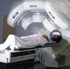
Radiosurgery Using the Linear Accelerator
Linear accelerator (LINAC) radiosurgery is similar to the Gamma Knife procedure and its four phases: head frame placement, imaging, computerized dose planning and radiation delivery. Unlike the Gamma Knife, which remains motionless during the procedure, part of the LINAC machine called a gantry rotates around the patient, delivering radiation beams from different angles. Compared to the Gamma Knife, the LINAC is able to use a larger x-ray beam, which enables it to treat larger tumors more uniformly and with less repositioning.
What will I feel during this procedure?
A nurse will place a small needle in your hand or arm to give medications, if needed, and a contrast material. Before the neurosurgeon positions and attaches your head frame, you will be injected with a local anesthetic in the front and back of your head to numb your scalp. These shots are only slightly uncomfortable and will help to minimize the discomfort of the head frame. As the head frame is pinned to your skull, you will feel pressure or tightness that typically disappears within 15 minutes.
Radiosurgery treatments are similar to having an x-ray. You will not be able to see, feel or hear the x-rays. There is no pain or discomfort from the actual treatment. If you experience pain for other reasons, such as back pain or discomfort from the head frame, you should let your doctor or nurse know.
When the head frame is removed, there may be some minor bleeding from the pin sites that will be bandaged. You may experience nausea and/or a headache and can ask for medication to help make you feel more comfortable.
Courtesy of: http://www.radiologyinfo.org/en/info.cfm?pg=stereotactic&bhcp=1